Case Studies
Examples of quantitative imaging analyses and biomarker outputs delivered across research and clinical development programs.
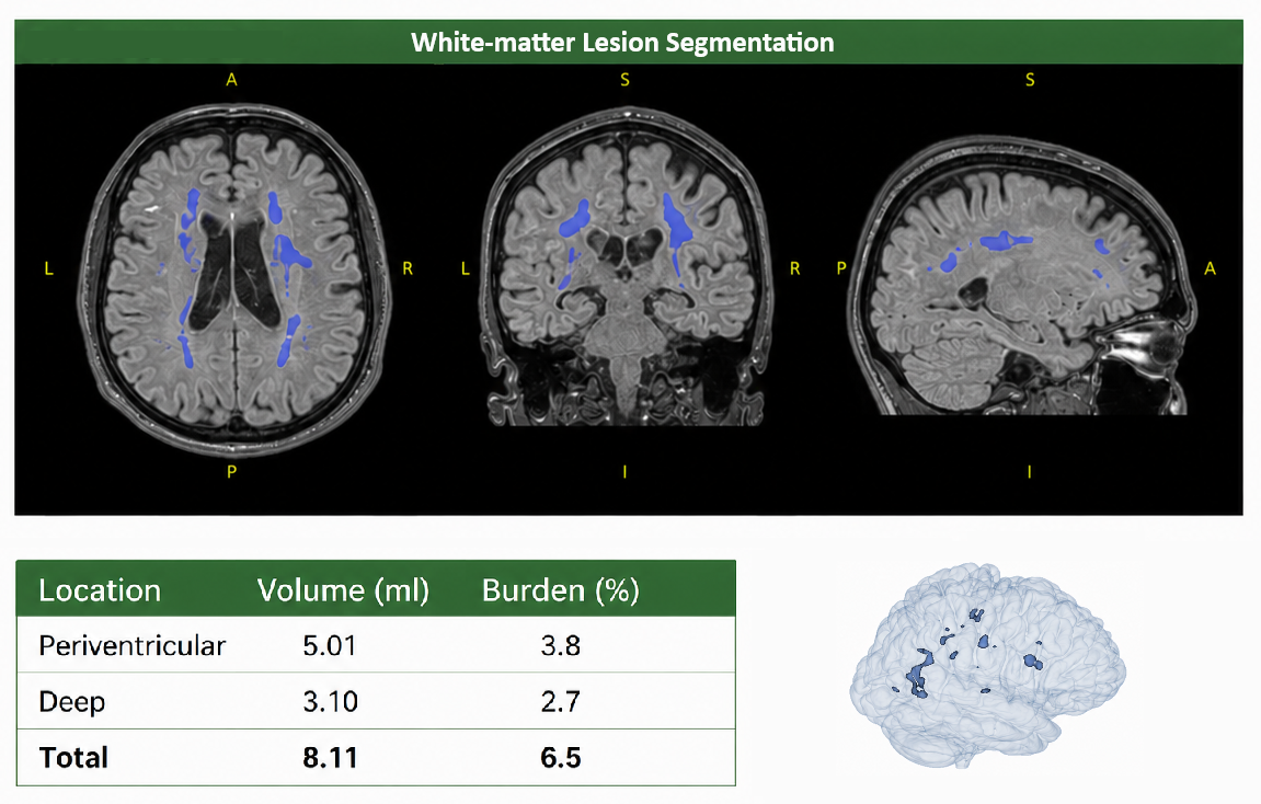
White-matter lesion load segmentation
Automated lesion segmentation with volumetric and burden quantification by anatomical location, alongside a 3D lesion map.
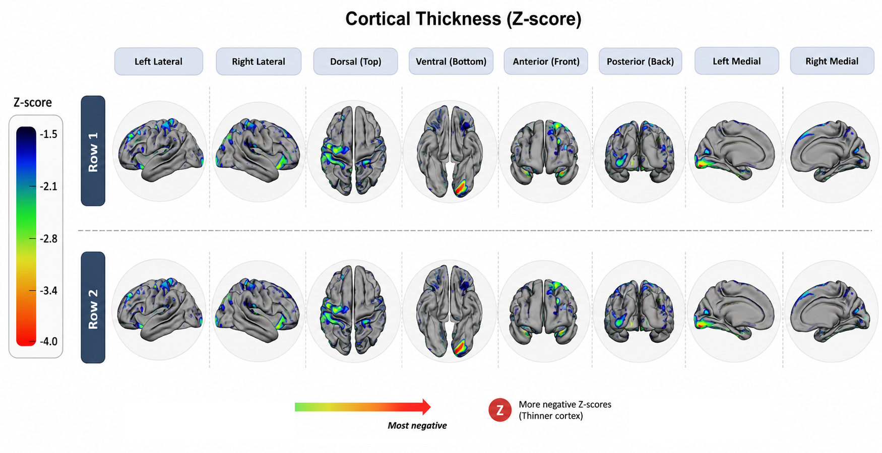
Cortical thickness estimation
Vertex-wise cortical thickness mapped across the cortical surface, with warmer colours indicating greater atrophy relative to normative reference values.
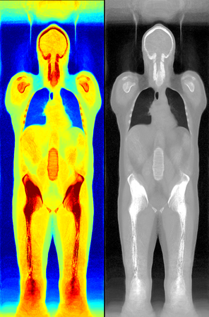
Whole-body registration template
A whole-body reference template used to intrinsically co-register longitudinal PET, CT, and MR studies for a given patient, ensuring scans from different time points and modalities align to a common anatomical space for consistent, side-by-side radiology reporting
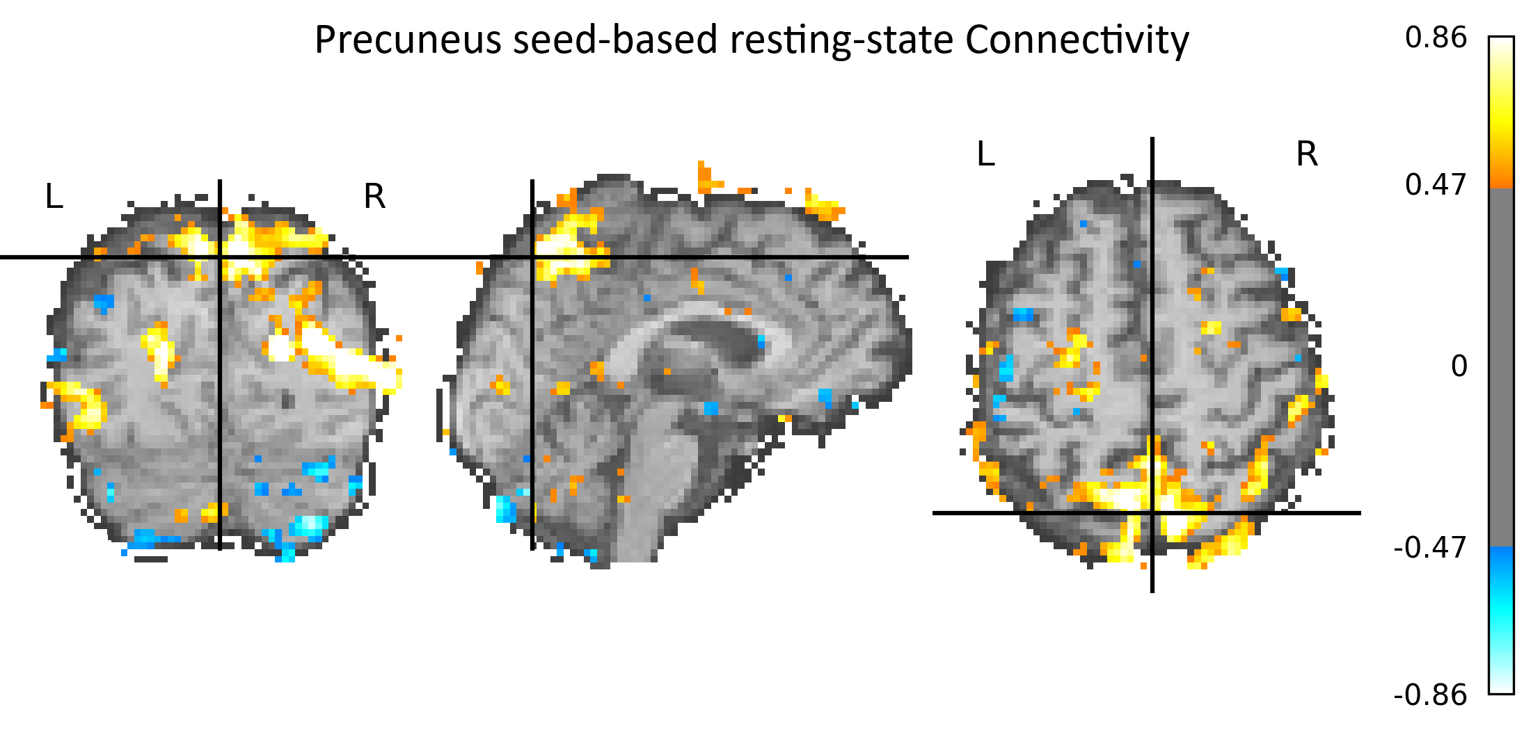
Resting-state fMRI connectivity
Precuneus seed-based resting-state connectivity, shown across axial, sagittal, and coronal slices, highlighting default-mode network coupling.
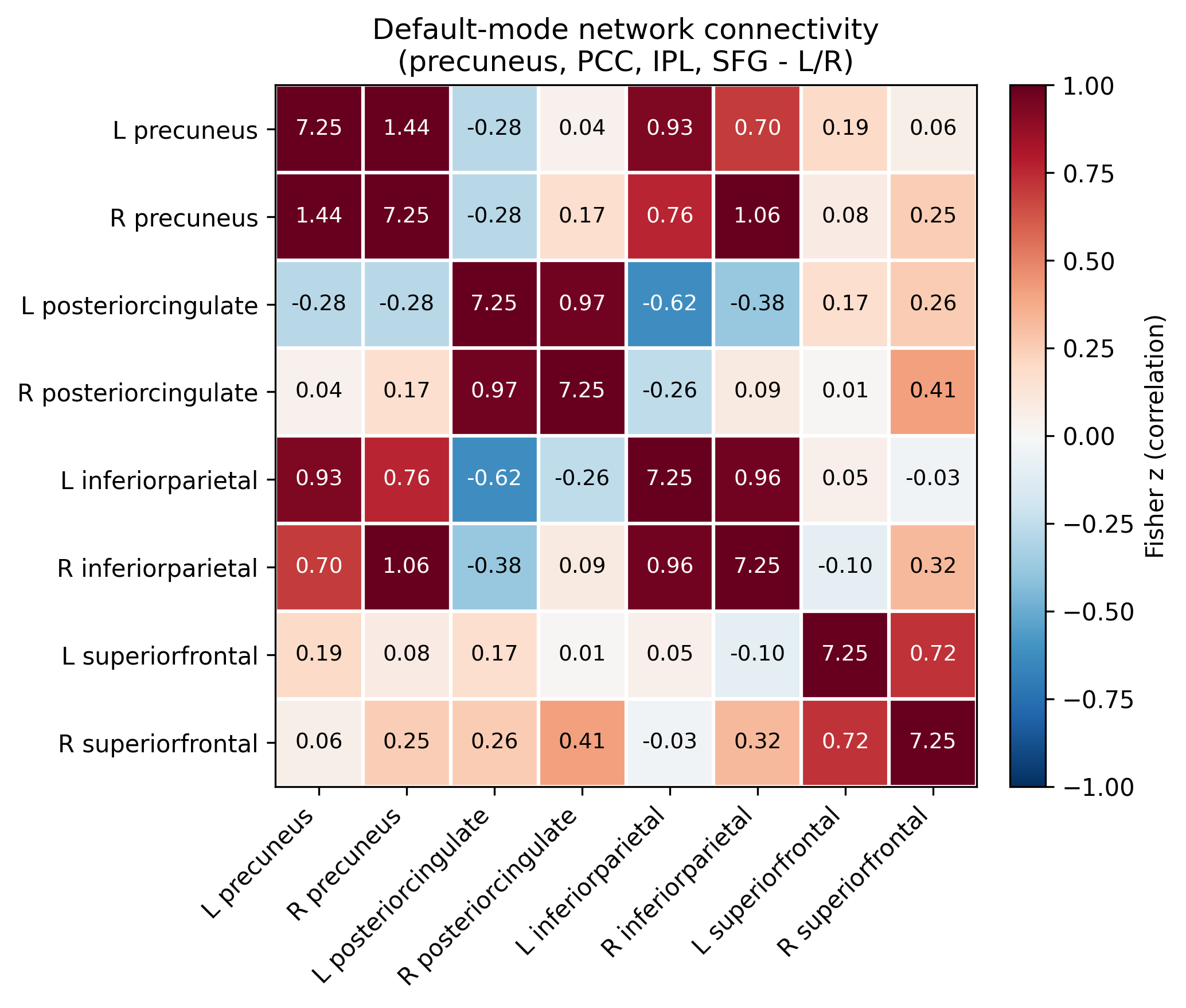
Default-mode network connectivity matrix
Region-to-region Fisher z-transformed correlation matrix across precuneus, posterior cingulate, inferior parietal, and superior frontal ROIs.